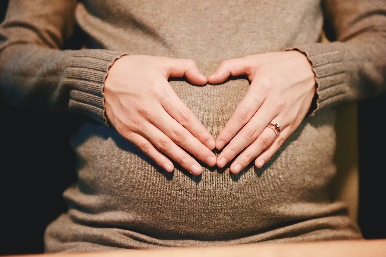
To understand changes in the brain during pregnancy, a neuroscientist chose to be regularly scanned with an MRI throughout her pregnancy. Dr. Elizabeth Chrastil, who co-authored a study of the findings was scanned three weeks before conceiving, during pregnancy, and for two years after her son was born in 2020.
The MRI scans and blood samples taken every few weeks revealed her brain underwent widespread reorganization. Some changes were short-lived and others lasted years. There are increases in plasma volume, metabolic rate, oxygen consumption and immune regulation. Much of the change is driven by the thousandfold surges in hormones such as estrogen and progesterone. Those hormones also reshape the central nervous system, especially areas of the brain that promote maternal behavior. Past studies have shown that the brain’s grey matter volume drops after the baby is born.
Grey matter is key in processing information, governing muscle control, sensory perception, and decision-making. These grey matter volume changes can still be seen up to 6 years after the mother delivers and remain traceable for decades. It’s possible the brain is preparing the mother by honing for example her vision and hearing so that she’s able to respond to her baby’s cries and needs.
Studies like these may help us predict outcomes such as post-partum depression and other pregnancy associated complications so we can target treatments for women who need it.
More Information
Scans capture sweeping reorganisation of brain in pregnancy
MRIs taken from before conception until two years after birth show some short-lived changes and some lasting years
Neuroanatomical changes observed over the course of a human pregnancy
Pregnancy is a period of profound hormonal and physiological changes experienced by millions of women annually, yet the neural changes unfolding in the maternal brain throughout gestation are not well studied in humans. Leveraging precision imaging, we mapped neuroanatomical changes in an individual from preconception through 2 years postpartum. Pronounced decreases in gray matter volume and cortical thickness were evident across the brain, standing in contrast to increases in white matter microstructural integrity, ventricle volume and cerebrospinal fluid, with few regions untouched by the transition to motherhood.
Unlocking the Maternal Brain: Groundbreaking Research Reveals Stunning Changes During Pregnancy
UC Irvine’s Charlie Dunlop School of Biological Sciences is proud to announce a new study published in Nature Neuroscience, revealing remarkable insights into the human brain during pregnancy. Conducted in collaboration with UC Irvine’s Associate Professor Elizabeth Chrastil, Ph.D., and UC Santa Barbara’s Associate Professor Emily Jacobs, Ph.D., this research presents the first comprehensive map of how a human brain undergoes neuroanatomical changes throughout pregnancy and beyond.